Eye Muscles
There are six extraocular muscles that control all of the movement of the eye. These muscles are the superior rectus, inferior rectus, lateral rectus, medial rectus, superior oblique, and inferior oblique. The muscles of the eye are designed to stabilize and move both eyes. Special nervous centers located throughout the brain and brainstem interact with each muscle pair (right and left) to coordinate precise movements of the eyes with limited conscious input.
All eye muscles have a resting muscle tone that is designed to stabilize eye position. During movements, certain muscles increase their activity while others decrease it. Eye movement has its own unique names. The movements of the eye include adduction (the moves in toward the nose); abduction (the moves out towards the ear); elevation (the eye moves up); depression (the eye moves down); intorsion (or incyclorotation; the top of the eye move rotates in towards the nose); and extorsion (or excyclorotation; the top of the eye move rotates in towards the ear).
What do the Eye Muscles Look Like?
Take a look at this image of the eye muscles. Each muscle has a primary and a secondary function. The image below is of the right eye (as if you were looking directly at the patient).
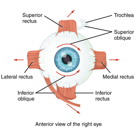
Superior Rectus
The superior rectus inserts at the anterior (front) portion of the eye, and its origin is behind the eye on the common ring tendon. Its primary function is to elevate the eye, and it has a mild secondary function of adduction and intorsion.
Inferior Rectus
The inferior rectus inserts at the anterior (front) portion of the eye, and its origin is behind the eye on the common ring tendon. Its primary function is to depress the eye, and it has a mild secondary function of adduction and extorsion.
Lateral Rectus
The lateral rectus inserts at the anterior (front) portion of the eye, and its origin is behind the eye on the greater wing of the sphenoid bone as well as the common ring tendon. Its primary function is to abduct the eye, and it has no secondary function.
Medial Rectus
The medial rectus inserts at the anterior (front) portion of the eye, and its origin is behind the eye on the common ring tendon. Its primary function is to adduct the eye, and it has no secondary function.
Superior Oblique
The superior oblique is unique. It inserts on the superior, lateral (ear-side), and posterior (back) of the eye. The anatomical origin is behind the eye on the lesser wing of the sphenoid bone, but the superior oblique muscle acts a pully, and loops back trough a connective tissue sling called the trochlea. Even though it is positioned above the eye, its unique use of the trochlea gives it a primary function is to intort the eye, and secondary functions of depression and abduction.
Inferior Oblique
The inferior oblique is also. It inserts on the inferior, posterior, lateral portion of the eye. Its origin is on the medial (middle) maxillary bone. Its primary function is extorsion, and its secondary functions are elevation and abduction.
How are the Eye Muscles Controlled?
The muscles that control eye movements are interesting. They use a larger supply of blood relative to their skeletal muscle counterparts, and only a few muscle fibers comprise a motor unit. This allows the cranial nerves responsible for coordinating eye movements to have precise control of the eyes.
Three cranial nerves are responsible for controlling the eye muscles. These are the third cranial nerve (oculomotor nerve), the fourth cranial nerve (trochlear nerve), and the sixth cranial nerve (abducens nerve). The secondary names of these nerves kind of give away what muscles they control. The fourth cranial nerve is named the trochlear nerve - it controls the superior oblique only. Similarly, the sixth cranial nerve (abducens nerve) controls only the lateral rectus - the primary abducting eye muscle. The third cranial nerve (oculomotor nerve) has a lot of responsibility. It controls the superior rectus, inferior rectus, medial rectus, and the inferior oblique muscles.
What is Eye Muscle Surgery?
Eye muscle surgery is commonly referred to as strabismus surgery. In some types of constant or even intermittent eye turns (strabismus), early surgery may be prescribed by an eye surgeon to increase the chance of returning or developing normal binocular vision.
Eye alignment surgery can restore normal appearance and is recognized as reconstructive surgery. There are many other benefits beyond restoring a normal appearance. Improved depth perception or complete reattainment of binocular vision, improved visual fields, eliminating or reducing double vision and improved social experiences since eye contact is hugely important in human communication and emotional responses.
It is essential to discuss the goals and expectations of the surgery with an ophthalmologist. During strabismus surgery, one or more of the eye muscles are moved to improve eye alignment. Strabismus surgery is usually performed as an outpatient procedure and does not require an overnight hospital stay.
References
(1) https://www.dartmouth.edu/~dons/part_1/chapter_4.html
Image: Extraocular muscles. Contributed by OpenStax, License: CC BY 4.0
The Vision Wiki
- Physiology of Vision
- The Brain
- Visual System
- Fifth Cranial Nerve
- Third Cranial Nerve
- Second Cranial Nerve
- Complex Cells in the Visual Cortex
- Sixth Cranial Nerve
- Seventh Cranial Nerve
- Visual Cortex
- Eye Muscles
- Binocular Neurons
- Simple Cells in the Visual Cortex
- Fourth Cranial Nerve
- Neuroplasticity
- Brain Injury
- Acquired Brain Injury
- Neuro Optometric Rehabilitation
- Traumatic Brain Injury
- Sports and Brain Injury
- The Eyes
- Gabor Patches
- Visual Snow
- Ocular Immune System
- Evolution Of Eyes
- Blue Field Entoptic Phenomena
- Bates Method
- Ophthalmology Lecture
- Optical Illusions
- Stereo Illusions
- Eye Problems
- Binocular Vision
- Lazy Eye
- Lazy Eye Treatments
- Reading
- Fields of Study
- Research
- Glaucoma
- Virtual Reality
- Organizations