Third Cranial Nerve
The third cranial nerve (CNIII) is also named the oculomotor nerve, as it primarily controls eye movement and eyelid movement (to a small degree). The portion of CNIII that controls eye and lid movement are called somatic fibers. CNIII also contains fibers from the autonomic nervous system (visceral or fight-or-flight system). These fibers affect the fine musculature of the iris (colored portion of the eye) by making the iris constrict or relax (to the observer, we see this as the pupil constricting or getting smaller or dilating or getting larger - the technical terms are miosis (constriction of pupil) and mydriasis (dilation of the pupil)). The autonomic fibers also affect the focusing or accommodation system of the eye.
Location and Course
All cranial nerves (all nerves for that matter) have a nucleus (cell body) and axon (that carries nerve impulses away from the nucleus to other structures). The nucleus of CNIII is located in the midbrain and is divided into six key parts. The axons from each portion of the nucleus extend anterior (moving towards the front of the head/face) and ultimately end in the superior rectus, inferior rectus, medial rectus, and inferior oblique muscles of the eye as well as the levator palpebrae muscle of the eyelid (somatic motor fibers). These subnuclei are aptly named the after what they innervate. All fibers from these nuclei serve the muscles on the same side of the eye - except for fibers from the superior rectus subnucleus. These fibers immediately cross over within the midbrain. All fibers from the midbrain and anterior now serve the same side of the body. The autonomic portion of CNIII originates from the Edinger-Westphal subnucleus, which supplies parasympathetic fibers to the iris sphincter and ciliary body muscles.
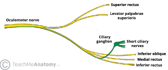
In its path forward, CNIII passes next to the posterior communicating artery (part of the Circle of Willis), through the top of the cavernous sinus, and through the superior orbital fissure. Within the cavernous sinus, CNIII interacts with the ophthalmic nerve (CNII) to receive sensory information. To further complicate things, CNIII then branches right after the superior orbital fissure. The superior branch of CNIII ends in the superior rectus muscle and levator palpebrae superioris. The inferior branch of CNIII ends in the medial rectus muscle, inferior rectus muscle, inferior oblique muscle, and ciliary ganglion. All of the fibers from CNIII remain on the right or left side - that is they do no decussate or cross over.
An example of the split is shown below, which shows the nerve just after leaving the superior orbital fissure.
A quick review of the location and destination of CNIII:
CNIII, superior portion
Starts in the MIDBRAIN and ends in the superior rectus of the eye and levator muscle of the eyelid.
CNIII, inferior portion
Starts in the MIDBRAIN and ends in the medial rectus, inferior rectus, inferior oblique, and ciliary ganglion on the SAME SIDE as its origin.
Oculomotor Nerve Problems
Damage to the oculomotor nerve can occur anywhere along its path. The clinical signs of damage can be helpful to localize where damage occurs.
Damage can cause the eye to move down and out on the affected side as the superior rectus, medial rectus, inferior rectus, and inferior oblique may become inactive. The down-and-out posture occurs due to unopposed action of the two remaining extraocular muscles, the superior oblique and lateral rectus. This acquired strabismus often causes significant diplopia (double vision).
Ptosis (eyelid droop) may occur as the levator no longer receives normal nerve impulses.
Dilation of the pupil may occur as the autonomic fibers are affected; this also often affects accommodation if the patient is young.
Root Atlas has a great set of videos that help explain how damage to CNIII affects a patient. Check it out below:
https://www.youtube.com/embed/videoseries?list=PLR8RWJD5FUMcYtkr44Kh-FkIXc5DxjdqU
Resources: https://medicine.yale.edu/cranialnerves/nerves/oculomotornerve/?tab=Clinical+correlation
The Vision Wiki
- Physiology of Vision
- The Brain
- Visual System
- Fifth Cranial Nerve
- Third Cranial Nerve
- Second Cranial Nerve
- Complex Cells in the Visual Cortex
- Sixth Cranial Nerve
- Seventh Cranial Nerve
- Visual Cortex
- Eye Muscles
- Binocular Neurons
- Simple Cells in the Visual Cortex
- Fourth Cranial Nerve
- Neuroplasticity
- Brain Injury
- Acquired Brain Injury
- Neuro Optometric Rehabilitation
- Traumatic Brain Injury
- Sports and Brain Injury
- The Eyes
- Gabor Patches
- Visual Snow
- Ocular Immune System
- Evolution Of Eyes
- Blue Field Entoptic Phenomena
- Bates Method
- Ophthalmology Lecture
- Optical Illusions
- Stereo Illusions
- Eye Problems
- Binocular Vision
- Lazy Eye
- Lazy Eye Treatments
- Reading
- Fields of Study
- Research
- Glaucoma
- Virtual Reality
- Organizations