What is Anomalous Retinal Correspondence?
Anomalous retinal correspondence (ARC) is a diagnosis often associated with strabismus (eye turn). To understand ARC, we need to first discuss how vision between both eyes works.
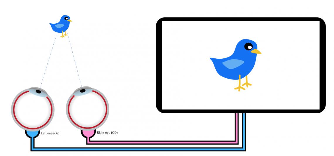
The eye contains over a million light-sensitive cells called photoreceptors. The two key types of photoreceptors are rods, which are important for low-resolution and movement detection, and cones, which are responsible for detail and high-resolution vision. The majority of cones are concentrated in a small area of each eye called the macula. When looking at something, the brain coordinates the movement of the two eyes so that the macula of each eye both point at the object of interest. The macula of each eye can be described as corresponding points in vision - each macula, when the eyes are properly aligned, both the right and left macula point at the same object in space. Now, picture on a broader scale each section of the retina of the right eye corresponding with a section on the retina of the left eye. Because the eyes have thousands and thousands of corresponding points between each eye, the brain can sort and combine images from both the right and left eye into one single image. Otherwise stated, correspondence between points in the retina of each eye is important for single binocular vision.

In this simple example, both eyes are directed towards the blue bird, and the brain interprets this as a single image of a blue bird.
Whew! So...what happens if the eyes don't point in the same direction, like in a patient with strabismus?
If one eye is turned in or out, this means that one of the eyes is not using the central part of the retina - the macula (and thus the part with the best vision) - to correspond (or match up with) the macula of the fellow eye. Now we have a problem - the patient will see double.
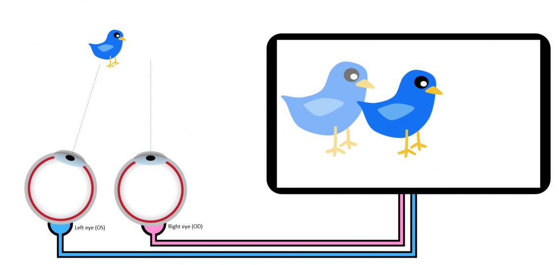
This is an example of strabismus or an eye turn below.

See the problem? The right eye is pointed away from the bird. The result is double vision (diplopia). If the patient is young, the visual system is quick to adapt - to prevent double vision, the brain "learns" to ignore the doubled image. This is called suppression (or more correctly, cortical suppression, as the actual suppression doesn't occur at the level of the eye, but rather within the visual cortex of the brain). The problem is solved...sort of. True, the patient won't have double vision, but now the visual system becomes dependent on vision from just one eye. This person wouldn't have depth perception or other eye teaming skills - at least not very good ones!
What About ARC?
We still didn't discuss what anomalous retinal correspondence is, but now you have a better idea of how the visual system normally works if a patient has strabismus. Sometimes the visual systems takes a different approach to double vision - instead of suppressing the doubled image, the visual system adapts to try to create single vision. In the case of ARC, some area OTHER THAN the macula of the deviated eye pairs with the macula of the normal eye. The visual system (brain) then interprets these two anomalous visual points from each retina as corresponding (hence the name - anomalous retinal correspondence).
How Do You Get ARC?
A few key requirements need to be met for ARC to develop:
- The strabismus must occur early in life (this means the visual system is still developing or "plastic" as in "neuroplasticity", so many patients have a history of an eye turn as a baby)
- The amount of eye deviation must be constant (if it's variable, the brain can't consistently match points on the deviating eye's retina with the macula of the normal eye)
- The amount of eye deviation must be small (small deviations are more consistent and often have better vision because they use some cones near central vision)
- The eye deviation must be unilateral (eye deviations that alternate won't cause ARC as it's easier for the brain to simply switch which eye is being used and suppress the deviating eye)
If the above criteria are met, the brain may adapts to eye misalignment and develops ARC to create single vision.
So, Is ARC BAD?
Remember, we haven't fixed the eye deviation, and now the brain taught itself to "fix" the problem of double vision by mismatching points between the two eyes. This is a better alternative than seeing double but doesn't fix the problem of the eye turn or poor binocularity. There may even be a greater chance that a patient DEVELOPS double vision if they have ARC and do eye exercises, have surgery to straighten the eyes or undergo vision therapy. Patients with ARC should carefully weigh the risks and benefits of any treatment with their eye care professional before starting any type of treatment for binocular vision problems.
How Do I Know if I Have ARC?
Your eye care provider can perform a number of different tests to screen for ARC. A simple test is to use a set of lenses that have small lines on them - this is called the Bagolini lens test. Your eye care professional may use other tests such as the Worth 4 Dot, stereopsis tests, prisms, and a cover test to check your eye alignment and visual skills. Sometimes a test called the after-image test is used. Similar to a bright camera flash or looking at a bright light, your eye doctor may use an after-image to understand how your visual system aligns the eyes and the level of correspondence between the two eyes.
Can ARC Be Treated?
Yes, but it's not always simple. Occlusion therapy, a tool called an amblyoscope, and even VR may be used to help treat ARC - but this should be done under the guidance of an eye doctor that is comfortable treating ARC. The core goal of ARC treatment depends on the patient - some patients are more interested in having eyes that point straight ahead, while others want to try to obtain the best vision possible in each eye. It's best to discuss this with your eye doctor to find out what the best option is for you!
The Vision Wiki
- Lazy Eye
- History of Lazy Eye
- Convergence Disorders
- Convergence Insufficiency
- Exophoria
- Esophoria
- Medical Terms
- Accommodation Disorders
- Accommodative Esotropia
- Accommodative Insufficiency
- Amblyopia
- Lazy Eye in Adults
- Strabismic Amblyopia
- Refractive Amblyopia
- Anisometropia
- Critical Period
- Strabismus
- Anomalous Retinal Correspondence
- Transient Strabismus
- Exotropia
- Esotropia
- Hypotropia
- Hypertropia
- Eye Problems
- Binocular Vision
- Physiology of Vision
- Lazy Eye Treatments
- Reading
- Fields of Study
- Research
- Glaucoma
- Virtual Reality
- Organizations
